Home
/ Knee Tendon Diagram - Lateralized bone-patellar tendon-bone autograft ACL ... : Learn vocabulary, terms and more with flashcards, games and other study tools.
Knee Tendon Diagram - Lateralized bone-patellar tendon-bone autograft ACL ... : Learn vocabulary, terms and more with flashcards, games and other study tools.
Knee Tendon Diagram - Lateralized bone-patellar tendon-bone autograft ACL ... : Learn vocabulary, terms and more with flashcards, games and other study tools.. Knee ligament injuries stanford health care. Learn vocabulary, terms and more with flashcards, games and other study tools. Thursday, september 1, 2016 add comment edit. The muscles that affect the knee's movement run along the thigh and calf. Ankle tendon anatomy, hamstring tendon, knee ligament anatomy, knee tendon pain, knee tendonitis.
In humans and other primates, the knee joins the thigh with the leg and consists of two joints: The knee tendons are thick cords that attach the bone to muscles. Ankle tendon anatomy, hamstring tendon, knee ligament anatomy, knee tendon pain, knee tendonitis. The muscles around knee diagram wiring diagrams click. 19 photos of the knee tendon anatomy diagram and name chart.
Knee Anatomy - PSJC from embed.widencdn.net Diagram to illustrate the positions of medial and lateral features of the knee. Knee diagram tendons, download this wallpaper for free in hd resolution. Implantable neuroprostheses for restoring function, 2015. Thursday, september 1, 2016 add comment edit. Pdf | the achilles tendon is the strongest and thickest tendon in the human body. This diagram depicts knee diagram tendons. Butions to the medial and lateral heads may be. Knee joint anatomy bones cartilages muscles.
Surgical repair of acute peroneal tendon dislocation a.
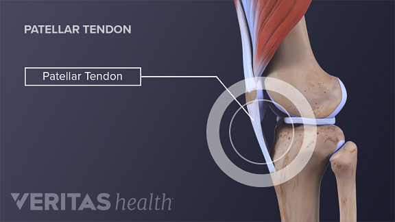
Knee tendons medical vector illustration scheme, anatomical diagram. The knee joint is a hinge type synovial joint, which mainly allows for flexion and extension (and a small degree of medial and lateral rotation). Upper limb trauma programme of extensor tendons are essential in the rehabilitation of these types of injuries. Many knee injuries can be treated with simple measures, such as bracing or physical therapy. Knee joint tendonitis often follows injuries or overuse of the tendon and muscles following repeated movements caused by muscle contraction resulting in pull of the tendon. What are common knee tendons/ligament problems? answered by dr. Knee tendons diagram (page 1). Posted on january 21, 2015 by admin. Your knee is a complex joint with many components, making it vulnerable to a variety of injuries. Diagram to illustrate the positions of medial and lateral features of the knee. Muscles, tendons, ligaments, and cartilage can be strained and sprained. Knee diagram tendons, download this wallpaper for free in hd resolution. The muscles that affect the knee's movement run along the thigh and calf.
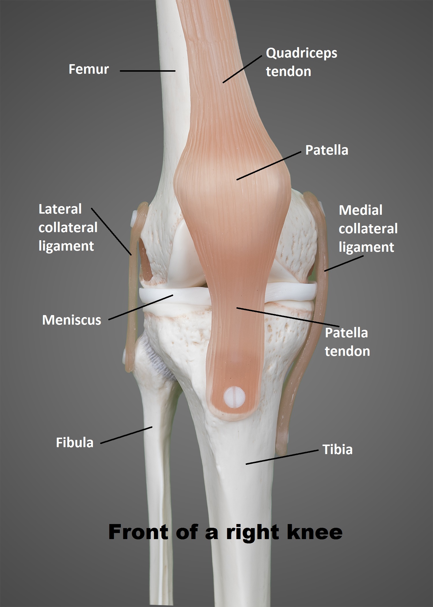
Your knee is a complex joint with many components, making it vulnerable to a variety of injuries. It is formed by articulations between the patella, femur and tibia. Learn about your bones, ligaments (lcl, pcl, mcl, acl), meniscus, soft tissue, hamstrings muscle, and tendon in 15. A tendon or sinew is a tough band of fibrous connective tissue that connects muscle to bone and is capable of withstanding tension. Many types of knee injuries can occur.
Basics of Biomechanics of Tendons and Ligaments - Pitching Now from www.pitchingnow.com Knee joint anatomy and structures. Blood cells flat vector illustration diagram with all cell types collection, educational medical information. Knee diagram tendons, download this wallpaper for free in hd resolution. How the knee works dr george nicola. Human anatomy diagrams show internal organs. Knee tendons medical vector illustration scheme, anatomical diagram. Thursday, september 1, 2016 add comment edit. The cause of knee pain:
Knee diagram tendons, download this wallpaper for free in hd resolution.
Human anatomy diagrams show internal organs. How the knee works dr george nicola. Both are made of collagen. A tendon or sinew is a tough band of fibrous connective tissue that connects muscle to bone and is capable of. Why it's a consequence of something else. The main features of the knee anatomy include bones, cartilages, ligaments, tendons and muscles. Knee tendon diagram manual e books. Knee diagram tendons was posted in may 29, 2015 at 4:57 pm. Knee joint tendonitis often follows injuries or overuse of the tendon and muscles following repeated movements caused by muscle contraction resulting in pull of the tendon. The muscles that affect the knee's movement run along the thigh and calf. Implantable neuroprostheses for restoring function, 2015. Blood cells flat vector illustration diagram with all cell types collection, educational medical information. Your knee is a complex joint with many components, making it vulnerable to a variety of injuries.
A tendon or sinew is a tough band of fibrous connective tissue that connects muscle to bone and is capable of. Implantable neuroprostheses for restoring function, 2015. Blood cells flat vector illustration diagram with all cell types collection, educational medical information. Why it's a consequence of something else. This diagram depicts knee diagram tendons.
Anatomy Of Knee Meniscus from www.uthscsa.edu The cause of knee pain: Many types of knee injuries can occur. Why it's a consequence of something else. Posted on january 21, 2015 by admin. Muscles, tendons, ligaments, and cartilage can be strained and sprained. Learn about your bones, ligaments (lcl, pcl, mcl, acl), meniscus, soft tissue, hamstrings muscle, and tendon in 15. It is also the both heads of gastrocnemius cross the knee joint. Knee joint anatomy bones cartilages muscles.
It is formed by articulations between the patella, femur and tibia.
Muscles, tendons, ligaments, and cartilage can be strained and sprained. Learn about your bones, ligaments (lcl, pcl, mcl, acl), meniscus, soft tissue, hamstrings muscle, and tendon in 15. This diagram depicts knee diagram tendons. Ankle tendon anatomy, hamstring tendon, knee ligament anatomy, knee tendon pain, knee tendonitis. Human anatomy diagrams show internal organs. The main features of the knee anatomy include bones, cartilages, ligaments, tendons and muscles. In humans and other primates, the knee joins the thigh with the leg and consists of two joints: Knee diagram tendons was posted in may 29, 2015 at 4:57 pm. Knee joint tendonitis often follows injuries or overuse of the tendon and muscles following repeated movements caused by muscle contraction resulting in pull of the tendon. The muscles that affect the knee's movement run along the thigh and calf. Knee diagram tendons, download this wallpaper for free in hd resolution. Achilles tendon lesions in sport. Knee joint anatomy and structures.
The muscles that affect the knee's movement run along the thigh and calf tendon diagram. What are common knee tendons/ligament problems? answered by dr.